A radiologist reviewing a complex tibial plateau fracture on a standard PACS viewer faces an immediate problem: the generic tools weren’t designed for orthopedic assessment. Measurements require manual workarounds, templating for implant sizing doesn’t exist, and comparing pre- and post-operative images means toggling between multiple windows. This friction costs time and, more critically, can compromise diagnostic accuracy. Orthopedic imaging demands specialized PACS tools that address the unique challenges of musculoskeletal assessment, from automated angle calculations to precise implant templating. Practices that recognize this distinction and invest in purpose-built solutions see measurable improvements in surgical planning accuracy and reduced revision rates. The difference between general radiology workflows and orthopedic-specific requirements isn’t subtle: it’s the difference between making educated guesses and having precise data that guides surgical decisions.
The Evolution of Orthopedic Imaging and PACS Integration
Transitioning from Traditional Viewers to Specialized Orthopedic PACS
The shift from film-based imaging to digital PACS represented a massive leap for radiology departments. Yet orthopedic surgeons quickly discovered that first-generation PACS platforms treated all imaging studies identically. A chest X-ray and a standing hip-to-ankle alignment study received the same viewing tools, despite radically different clinical needs.
Modern orthopedic PACS platforms address this gap with specialty-specific functionality. OmniPACS and similar cloud-based solutions now offer measurement tools calibrated for bone and joint assessment, hanging protocols designed for orthopedic comparison studies, and integration with digital templating systems that surgeons actually use.
Key Differences Between General and Orthopedic-Specific Workflows
General radiology workflows prioritize throughput and standardized reporting. Orthopedic workflows demand interactive measurement capabilities, the ability to overlay implant templates at true scale, and tools for tracking deformity correction over time.
A spine surgeon reviewing scoliosis progression needs instant Cobb angle measurements across multiple studies. A joint replacement specialist requires templating that accounts for magnification factors and patient anatomy. These aren’t optional features: they’re fundamental requirements that separate adequate imaging from imaging that directly improves surgical outcomes.
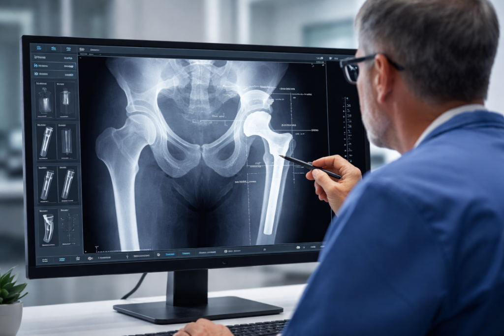
Advanced Digital Templating and Pre-operative Planning
Automated Sizing for Joint Replacement Implants
Pre-operative templating has evolved from acetate overlays on lightboxes to sophisticated digital systems integrated directly into PACS viewers. Modern platforms automatically calibrate for magnification using reference markers, then allow surgeons to overlay manufacturer-specific implant templates at true scale.
The accuracy gains are substantial. Studies consistently show that digital templating predicts the correct implant size within one size in over 85% of cases, compared to roughly 65% with manual methods. This precision reduces intraoperative decision-making, shortens surgical time, and decreases the inventory of implants that must be sterilized and available.
Reducing Intraoperative Surprises Through Precise Measurements
When templating fails, surgeons face difficult choices mid-procedure. An undersized femoral component discovered after the femoral canal is prepared creates delays while additional sizes are opened or emergency orders are placed. Oversized components risk fracture during impaction.
Orthopedic PACS tools that support accurate templating eliminate most of these scenarios. The combination of calibrated imaging, manufacturer template libraries, and measurement verification creates a surgical plan that holds up in the operating room.
Specialized Measurement Tools for Musculoskeletal Assessment
Automated Cobb Angle and Spinal Alignment Calculations
Manual Cobb angle measurement requires identifying the end vertebrae, drawing lines along the endplates, and calculating the angle between the perpendicular lines. This process introduces inter-observer variability of 3–5 degrees, enough to change treatment recommendations.
Automated measurement tools reduce this variability to under 2 degrees while completing calculations in seconds rather than minutes. For practices that have monitored adolescent idiopathic scoliosis patients over the years, consistent measurements are essential to distinguish true progression from measurement error.
Leg Length Discrepancy and Limb Deformity Analysis
Standing hip-to-ankle radiographs require precise measurement protocols that general PACS viewers don’t support natively. Orthopedic-specific tools provide templates for mechanical axis determination, anatomical axis measurement, and leg length calculation with sub-millimeter precision when properly calibrated.
OmniPACS offers these specialized measurement capabilities through its cloud-based platform, enabling surgeons to perform complex deformity analysis from any location with consistent results.
Enhancing Diagnostic Clarity with Image Manipulation Features
Multi-planar Reconstruction (MPR) for Complex Fractures
CT imaging of complex fractures produces hundreds of axial slices that must be reconstructed for surgical planning. Multi-planar reconstruction tools allow surgeons to visualize fracture patterns in coronal, sagittal, and oblique planes without returning to the CT console.
For tibial plateau fractures, acetabular fractures, and complex periarticular injuries, MPR capabilities transform surgical planning. Surgeons can identify fracture lines, measure fragment displacement, and plan surgical approaches using reconstructions optimized for their specific questions.
Side-by-Side Comparison for Longitudinal Patient Tracking
Orthopedic care frequently spans months or years, with imaging studies at multiple time points. Effective comparison requires more than placing two images adjacent: it demands registration tools that align anatomy, measurement persistence that carries forward previous calculations, and display protocols that present relevant comparison studies automatically.
Practices tracking fracture healing, fusion progression, or implant stability need PACS systems that enable effortless longitudinal comparisons, rather than requiring manual study retrieval and window setup.
The Role of AI and Automation in Reducing Human Error
AI-Driven Fracture Detection and Prioritization
Artificial intelligence applications in orthopedic imaging have moved beyond research into clinical deployment. Current AI tools detect fractures with sensitivity exceeding 90% across common injury patterns, flag studies requiring urgent attention, and reduce the risk of missed findings.
These tools work best as a safety net rather than a replacement for radiologist interpretation. When AI identifies a potential fracture that the initial reader missed, the system can trigger a secondary review before the patient leaves the facility.
Standardizing Reports to Improve Communication with Surgeons
Structured reporting templates ensure that orthopedic imaging reports consistently address the clinical questions surgeons need answered. Rather than free-text descriptions that vary by radiologist, standardized reports include specific measurements, classifications, and surgical planning information in predictable locations.
This standardization reduces phone calls between surgeons and radiologists, decreases the risk of critical findings being buried in report text, and creates data that practices can analyze for quality improvement.
Future-Proofing Orthopedic Practices Through Cloud-Based PACS
The infrastructure demands of modern orthopedic imaging continue to grow. Higher-resolution imaging, 3D reconstructions, and AI processing require computational resources that strain on-premise systems. Cloud-based PACS platforms address these challenges by shifting storage and processing to scalable infrastructure.
OmniPACS exemplifies this approach, providing orthopedic practices with enterprise-level imaging capabilities without the capital investment and IT burden of traditional systems. Multi-location practices gain consistent access to imaging studies regardless of where they were acquired, while automatic updates ensure the platform incorporates new features without manual intervention.
For practices evaluating their imaging infrastructure, the question isn’t whether to adopt specialized orthopedic tools: it’s whether their current platform supports the diagnostic accuracy their patients deserve.
Frequently Asked Questions
What makes orthopedic PACS different from standard radiology PACS?
Orthopedic PACS platforms include specialized measurement tools for angles, alignment, and leg length, digital templating for implant sizing, and display protocols optimized for musculoskeletal comparison studies. These features address workflows that general PACS systems don’t support natively.
How accurate is digital templating for joint replacement surgery?
Digital templating predicts the correct implant size within one size in approximately 85–90% of cases when properly calibrated. This accuracy depends on consistent imaging protocols and appropriate magnification markers.
Can cloud-based PACS handle the large file sizes from orthopedic CT and MRI studies?
Modern cloud platforms handle large imaging studies effectively, with compression algorithms and progressive loading that make remote access practical even for complex CT reconstructions. However, sufficient internet bandwidth remains necessary for seamless real-time performance, even with optimization techniques.
Does AI fracture detection replace radiologist interpretation?
AI tools serve as a safety net rather than a replacement for trained radiologists. They excel at flagging potential findings for secondary review and prioritizing urgent studies, but final interpretation remains with qualified physicians.
Building a Foundation for Better Orthopedic Outcomes
The tools available for orthopedic imaging assessment have matured significantly, and practices that invest in specialized PACS capabilities gain measurable advantages in diagnostic accuracy and surgical planning precision. From automated measurements that reduce variability to templating systems that predict implant sizing, these features directly impact patient outcomes.For practices seeking a modern approach to medical imaging, OmniPACS offers cloud-based solutions tailored to orthopedic workflows, without the complexity of traditional systems. Explore how OmniPACS can support your practice.